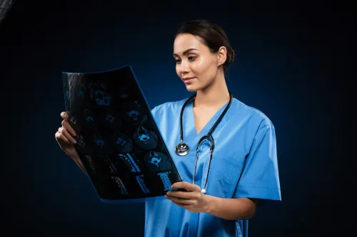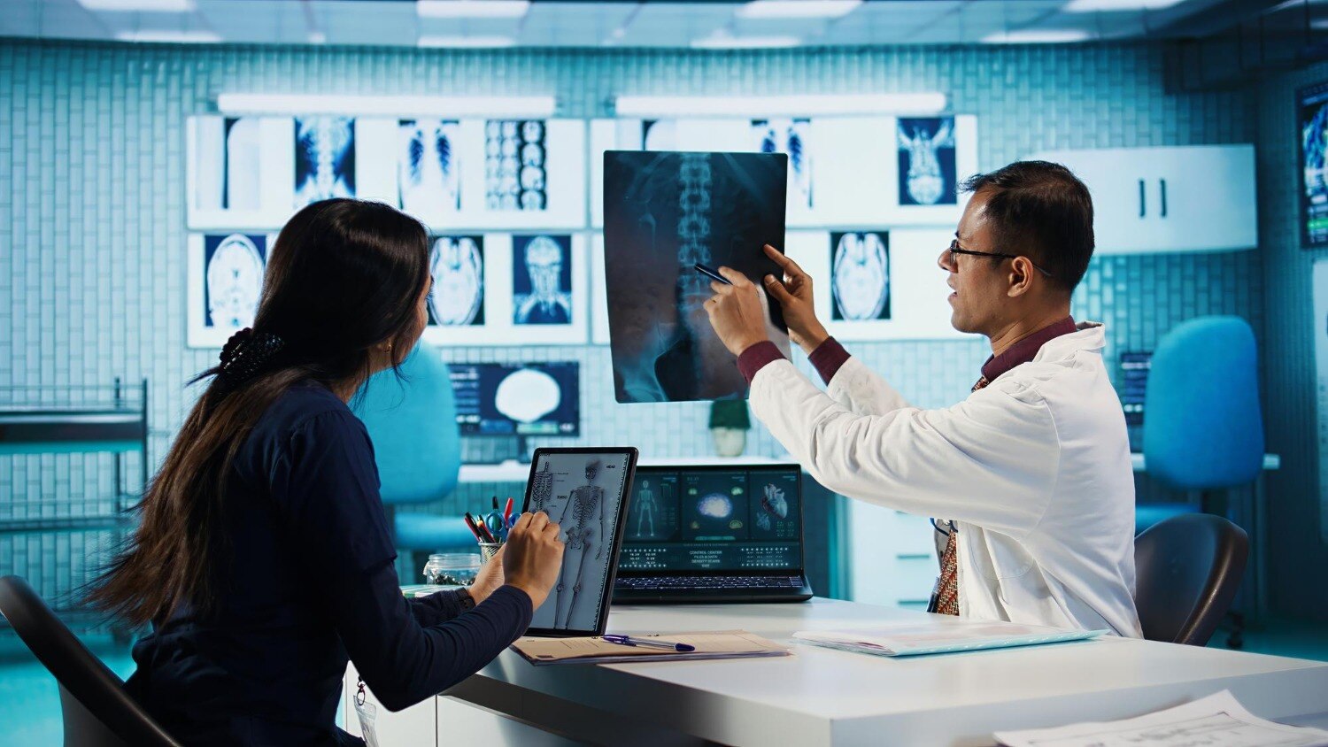
A radiologist’s report is only as reliable as the images and patient information entering the reporting workflow. By following a hospital X-ray from request verification through positioning, exposure, quality review and image transfer, you can understand where technicians protect accuracy, safety and reporting speed—and what training prepares someone to perform that work.
Key takeaways
- Confirm patient identity, body part and clinical question before imaging.
- Use immobilisation, modified positioning and clear instructions for trauma patients.
- Check warning lights, exposure controls, accessories and protective barriers before use.
- Verify accreditation, clinical training, eligibility and placement details before applying in Pune.
What happens during an X-ray technician’s hospital shift?
A doctor’s request becomes a usable study only after the technician confirms that the correct patient is being examined for the correct clinical question. The technician produces images; the radiologist interprets them and issues the report.
- Reviews the request, checking the examination, body region, symptoms and requested views.
- Confirms the patient’s name, date of birth and identity band before positioning.
- Explains the procedure, checks relevant pregnancy information for abdominal or pelvic imaging, and answers safety questions.
- Positions the patient and selects the required projections, such as posteroanterior and lateral views for a chest examination.
- Sets exposure factors, collimates the beam and operates the X-ray system to obtain each image.
- Checks positioning, coverage, sharpness, exposure and visible anatomy before releasing the patient.
- Repeats an image only when necessary, because an avoidable repeat adds radiation without improving diagnosis.
- Records the examination and sends the approved images through the hospital’s image-archiving system to the radiologist.
These x ray technician duties support diagnosis without replacing it. A technician can alert the supervising team to an urgent workflow or safety concern, but must not label an image as a fracture, pneumonia or another diagnosis.
For contrast-enhanced CT or fluoroscopy, the technician may prepare equipment and the patient. Screening, contrast administration and reaction management follow hospital protocol and the authorised medical team’s direction.
Which examinations and imaging departments involve X-ray technicians?
Plain radiography covers the common chest, abdominal, extremity, spine and trauma studies performed with a fixed or mobile X-ray unit. The technician confirms the request, positions the patient, selects views such as posteroanterior, anteroposterior, lateral or oblique, operates the equipment, checks image quality and sends the images for interpretation.
The radiologist reports the findings; the technician does not diagnose.
Common radiographs include:
- Chest X-ray for lungs, heart size and acute chest symptoms
- Abdominal X-ray for bowel gas patterns, obstruction or swallowed objects
- Extremity views of the fingers, wrist, ankle, knee or shoulder
- Spine views of the cervical, thoracic or lumbar region
- Trauma and mobile radiographs for patients who cannot reach the imaging room
Radiology technician applications can extend into other departments, but each modality changes the equipment, safety duties and training required.
| Modality | Technician’s role | Key difference |
|---|---|---|
| Plain X-ray | Positions patients and acquires single or multiple projections | Uses ionising radiation for 2D images |
| CT | Operates a rotating X-ray scanner and prepares patients under protocol | Produces cross-sectional images; contrast work needs authorised medical supervision |
| Fluoroscopy | Supports real-time X-ray examinations | Requires additional radiation and procedure-room training |
| Mammography | Performs specialised breast projections | Uses dedicated equipment and compression technique |
| MRI | Requires separate magnetic-field safety training | Uses no ionising radiation |
| Ultrasound | Usually performed by a sonographer | Uses sound waves, not X-rays |
How do technicians adapt radiography for trauma and difficult patients?
An immobile patient does not make a diagnostic radiograph impossible; it changes the projection, support and equipment. The radiographer checks the request, identity, injury precautions, lines and tubes, then coordinates with emergency staff before moving the patient or positioning the detector.
- Chest: A portable anteroposterior (AP) image can be taken with the patient supine or semi-upright. The technician places the detector behind the back, centres it over the chest and gives breathing instructions only if the patient can follow them.
- Abdomen: A supine AP image avoids standing. If free air or fluid is suspected, a horizontal-beam lateral or decubitus view can show it without requiring the patient to stand.
- Extremity: The injured arm or leg is supported in its existing position. The technician changes the tube angle or uses a horizontal beam instead of rotating the limb, then obtains additional views that do not worsen pain or injury.
- Spine: A cross-table lateral view can image the cervical or thoracic spine while the patient remains immobilised. Staff maintain spinal precautions; the technician does not remove a collar or ask for rotation without clinical clearance.
- Major trauma: Portable images are acquired around splints, dressings and monitoring equipment. The team controls nearby people, cables and infection risks before exposure.
The technician adjusts collimation, exposure and positioning to include the required anatomy while limiting motion and artefacts. If a textbook position is unsafe, a technically limited image is documented rather than repeated unnecessarily. The radiologist interprets the images; the technician sends usable views and flags workflow or safety concerns.
How do equipment checks and radiation safety protect patients?
Before an exposure, the technician confirms the patient’s identity, body part and clinical question, then checks whether the examination is justified. A duplicate request or an available recent image can prevent unnecessary dose. The technician applies ALARA—keeping exposure as low as reasonably achievable while preserving diagnostic quality—not by lowering settings until the image becomes unusable.
Equipment checks include detector condition, cables, warning lights, exposure controls and scheduled quality-assurance tests. In Pune hospitals, AERB requirements cover X-ray equipment, room shielding, operating procedures and personnel monitoring. The technician also asks relevant pregnancy questions and documents concerns; a possible pregnancy prompts a risk-benefit discussion with the authorised clinician, not an automatic cancellation.
Use this safety checklist before and during acquisition:
- Collimate to the requested anatomy. A smaller field reduces irradiated tissue and scatter.
- Select exposure factors and projections that answer the clinical question with the fewest images.
- Ask staff and visitors to leave the room. If help is essential, place the helper outside the primary beam, maximise distance and provide protective apparel.
- Use fixed barriers or room shielding; a lead apron cannot correct poor collimation or an unjustified examination.
- Wear and correctly position the assigned dosimeter. It records occupational exposure; it does not protect the patient.
- Review anatomy coverage, positioning, motion, exposure, artefacts and markers before releasing images. A blurred or incomplete image can force a repeat.
- Record exposure details and escalate equipment faults or safety concerns. The radiologist interprets the images and issues the report.
What should you check before applying for X-ray training in Pune?
A course title does not prove that you will handle patients safely. For x ray for technician course applications in pune, ask for written details on hospital postings, supervised hours, assessment methods and the qualification’s awarding body.
Compare the route against these points:
- Clinical rotation: Ask whether placements include outpatient radiography, emergency imaging, portable X-ray, trauma cases and night-shift exposure.
- Equipment access: Check how many rooms students use and whether training includes computed radiography, digital radiography, mobile units, image archiving and radiation dosimeters.
- Supervision: Confirm who observes positioning, exposure selection, patient identification, pregnancy screening and image-quality checks. A classroom certificate cannot demonstrate these skills.
- Recognition: Request the exact certificate, diploma or degree name, awarding institution and internship record. Do not rely on “radiology” in the course title.
| Route | What to verify | Limitation |
|---|---|---|
| x ray technician course in pune | Plain radiography, positioning, radiation safety and documented clinical practice | It may not qualify you for CT, MRI or fluoroscopy |
| medical imaging course in pune | Covered modalities, posting hours and access to CT, mobile imaging or fluoroscopy | Broader coverage can mean less depth in plain X-ray |
| Degree-level radiography route | University recognition, eligibility, clinical syllabus and supervised competencies | It requires more time and formal entry requirements |
K. P. Paramedical Institute is worth comparing on the same evidence: ask for its clinical training arrangement, practical assessment records and qualification recognition rather than choosing from the course name alone. MRI requires separate magnetic-field safety training, while contrast work requires hospital protocol and authorised supervision.
Related products
 | Best X-RAY The X-Ray Technician Course at K P Paramedical Institute is a career-oriented paramedical program designed to prepare students for careers in... View product → |
 | Best DRIT The Diploma in Radiology & Imaging Technology (DRIT) at K P Paramedical Institute is a career-oriented paramedical program designed to prepare... View product → |
Frequently asked questions
What happens during an X-ray technician’s hospital shift?
A technician reviews the request, confirms the patient and examination, prepares the equipment, positions the patient, captures images and records the study for radiologist review.
Which examinations and imaging departments involve X-ray technicians?
Technicians support plain radiography such as chest, limb, spine and abdominal examinations, along with portable imaging, emergency radiography and specialised departments that use X-ray equipment.
How do technicians adapt radiography for trauma and difficult patients?
They use safe positioning changes, immobilisation aids, portable equipment and concise instructions while avoiding unnecessary movement of an injured patient.
How do equipment checks and radiation safety protect patients?
Technicians check the machine and room before use, confirm exposure settings, collimate the beam, use shielding when appropriate and apply the minimum exposure needed for a diagnostic image.
What should you check before applying for X-ray training in Pune?
Check the course level, eligibility, syllabus, practical hospital training, equipment access, assessment method, approval details and the institute’s written progression or placement information.