Students comparing healthcare courses need more than a promise of career opportunity: they need to know which technical skills an X-ray programme builds, how much supervised practice it provides, and what work the qualification actually permits. By the end, you will be able to judge the practical benefits, compare course routes, assess local convenience against training quality, and question an institute before enrolling.
Key takeaways
- Practise patient identification, positioning, radiation safety and equipment handling.
- Confirm supervised clinical hours and access to functioning X-ray equipment.
- Match certificate, diploma or degree study to your intended healthcare role.
- Ask for recognition details, assessment methods and placement arrangements before enrolling.
Which practical skills make X-ray training useful at entry level?
For students, x ray technician course benefits begin with a usable technical foundation: recognising anatomy, confirming patient identity, explaining the examination and handling patients respectfully. The benefits of X-ray training in Daund depend on practising these skills on actual equipment under supervision, not memorising theory alone.
| Skill | Entry-level application | Why it matters |
|---|---|---|
| Anatomy recognition | Identify chest, joints and limb landmarks | Supports correct projections |
| Patient identification | Match the patient with the request | Prevents wrong-patient exposures |
| Communication | Explain instructions and check comfort | Reduces movement and anxiety |
| Positioning | Perform chest and limb projections | Places anatomy correctly |
| Exposure selection | Choose kVp and mAs | Controls penetration and dose |
| Image checks | Review coverage, rotation and artefacts | Prevents avoidable repeats |
| Infection control | Clean contact surfaces and use precautions | Limits cross-contamination |
| Emergency awareness | Recognise distress and seek help | Protects an unstable patient |
Accurate positioning and exposure selection reduce motion artefacts, unnecessary repeat exposures and delays for the reporting doctor. Training demands anatomy and physics study, repeated positioning practice, patient handling, standing during procedures and strict attention to instructions. The technician produces technically useful images; the technician does not independently diagnose them or issue radiology reports.
How does classroom learning become safe clinical competence?
A medical imaging course in Daund becomes clinical competence through supervised practice, not theory alone. Students follow the department’s workflow while learning to use fixed radiography units, mobile X-ray machines, image receptors, computed radiography cassettes, digital radiography detectors and exposure-control consoles.
- Receive the referral, identify the patient with approved identifiers and explain the examination.
- Check the examination request, projection, clinical question and relevant patient conditions.
- Position the patient and receptor, then select exposure factors such as kVp and mAs.
- Make the exposure under authorised supervision.
- Review positioning, collimation, exposure and motion on the image.
- Repeat an image only when a documented technical reason justifies it.
- Record the examination and send images through PACS using the DICOM workflow.
Radiology training in Daund should teach ALARA: keep exposure as low as reasonably achievable through reduced time, greater distance and effective shielding. Students use lead aprons, thyroid shields, collimation, structural barriers, controlled areas and personal dosimeters; shielding alone is not enough.
They practise communication and safe positioning for children, pregnancy assessment under facility policy, and patients who cannot remain still. Immobilisation choices can prevent motion and repeat exposures. Students must never operate X-ray equipment without authorised supervision.
Which training route fits your intended healthcare role?
For students comparing x ray technician course benefits, the choice depends on the role you want and the qualification an employer recognises. A short certificate may suit foundational plain X-ray work; a diploma may provide broader study of radiography, digital imaging and related modalities. The title alone proves neither.
| Route | Qualification and duration | Eligibility, practical scope and progression |
|---|---|---|
| X-ray technician certificate or short course | Entry-level; duration varies | Confirm school-leaving requirements, plain X-ray syllabus, documented practical hours and supervised equipment use. Progression may require further experience or higher study. |
| Diploma in Radiology & Imaging Technology | Higher-level; duration varies | Check eligibility, clinical placements, practical-hour allocation, digital radiography, image processing, PACS, DICOM and related modalities. It may support broader progression. |
Ask for the written syllabus, entry requirements, total duration, practical-hour log and awarding authority before enrolling. A medical imaging course in daund should be judged by these documents, not by “radiology” in its name.
Choose imaging if you enjoy anatomy, physics, equipment and patient positioning. ECG training centres on cardiac recordings, medical laboratory training on specimens, dialysis training on renal support, and operation-theatre training on surgical assistance.
CT, MRI and independent image interpretation require suitable higher qualifications and supervised experience. An X-ray technician produces technically useful images but does not diagnose them or issue radiology reports.
What should you verify before choosing an X-ray course in Daund?
Studying in Daund can reduce commuting pressure and make local access easier when you must balance travel, family responsibilities and clinical attendance. Those are real benefits of X-ray training in Daund, but convenience does not prove training quality or guarantee nearby employment.
Ask K. P. Paramedical Institute, or any provider you compare, for these records before paying:
| Question | Verify | Why it matters |
|---|---|---|
| Recognition | Current affiliation or approval, awarding body, examination authority and eligibility documents | Confirms what the qualification represents |
| Course structure | Syllabus, course duration and written total-fee schedule | Prevents title, time and cost surprises |
| Practical training | Lab timetable, equipment list, practical-hours allocation, faculty supervision and practical logbook format | Shows whether practice is documented and supervised |
| Clinical exposure | Clinical-training location, internship terms and safety protocol | Reveals where and how patient work occurs |
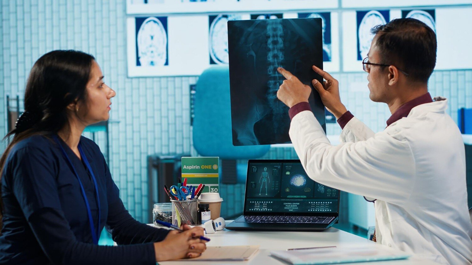
Do not accept demonstrations as clinical training. Check that students use actual fixed or mobile X-ray equipment, digital detectors or cassettes under authorised supervision, practise positioning and exposure selection, review images and record procedures.
Ask whether the qualification meets registration or employment requirements applicable to your intended Maharashtra role. Confirm this in writing with the relevant employer or authority, because course titles alone do not settle eligibility.
Ignore “best” claims, vague placement language and unverified employer lists. A nearby course is worth choosing only when its recognition, practical access and clinical arrangements withstand these checks.
Related products
 | Best X-RAY The X-Ray Technician Course at K P Paramedical Institute is a career-oriented paramedical program designed to prepare students for careers in... View product → |
 | Best DRIT The Diploma in Radiology & Imaging Technology (DRIT) at K P Paramedical Institute is a career-oriented paramedical program designed to prepare... View product → |
What work can the qualification lead to, and what comes next?
The qualification can support applications for entry-level technician work in hospitals, diagnostic centres, trauma units, outpatient clinics and mobile imaging services. These x ray technician course benefits depend on a recognised qualification, practical competence, registration rules, available vacancies and your willingness to work shifts or travel to nearby facilities.
Radiology training in Daund builds a technical foundation, not a job guarantee.
A technician operates imaging equipment, positions patients and checks image quality; a radiologist is the physician who interprets images and issues reports. A short certificate does not qualify you for every radiology role, and digital imaging does not make radiation harmless: exposure creep can produce an acceptable-looking image while increasing patient dose.
Confirm the course’s eligibility for the role you want.
After foundational training, realistic next steps include:
- Further supervised experience in plain X-ray, with documented practice and quality checks.
- A Diploma in Radiology & Imaging Technology, if you meet the institution’s eligibility requirements.
- Eligible study in CT, MRI or related specialities after meeting the relevant admission and supervision requirements.
Independent image interpretation and advanced-modality roles require a different qualification pathway. Compare the course level, clinical hours and progression rules before treating a certificate as equivalent to a diploma or degree.
Frequently asked questions
Which practical skills make X-ray training useful at entry level?
Useful entry-level skills include confirming patient identity, recognising basic anatomy, explaining examinations, positioning patients, handling equipment and following radiation-safety procedures.
How does classroom learning become safe clinical competence?
Students build competence through demonstrations, supervised practice on working equipment, patient communication, image-quality checks and feedback from qualified trainers.
Which training route fits your intended healthcare role?
A certificate may suit an introductory support role, while a diploma or undergraduate programme provides broader study for technician and allied-health pathways. Check the role and employer requirements first.
What should you verify before choosing an X-ray course in Daund?
Verify the institute's recognition, syllabus, trainer qualifications, equipment access, supervised clinical hours, assessment process, safety instruction and placement arrangements.
What work can the qualification lead to, and what comes next?
Training can support entry-level work in diagnostic imaging and clinical support. Next steps include checking local employer requirements, gaining supervised experience and pursuing further study.